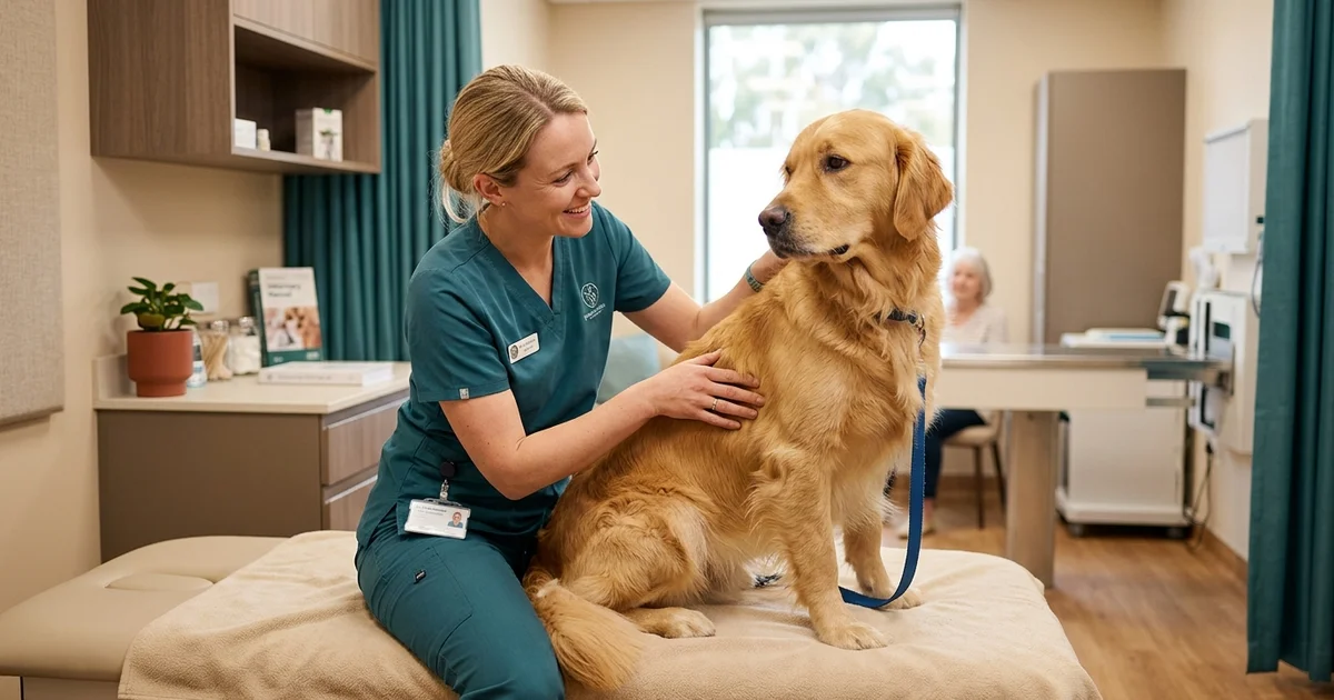
What the surgery actually involves
Canine mass and tumor removal is the surgical excision of a lump from your dog’s skin or the tissue beneath it. It covers everything from a small superficial growth to a deep subcutaneous mass sitting against muscle, and it is the most common planned surgery performed on dogs in the Brandywine Valley.
The operation is conceptually simple: the mass comes out and the resulting gap is closed. What separates a good result from a mediocre one is the planning that happens before the first incision, which is exactly what our mass and tumor removal service is built around.
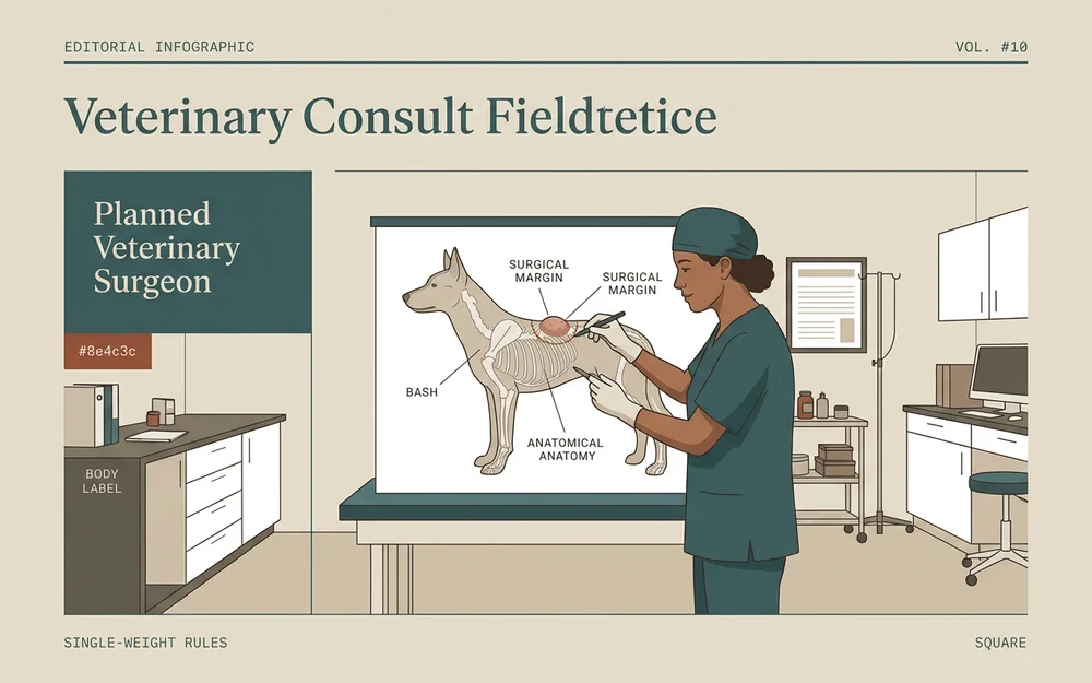
Excision versus biopsy
These two words get used interchangeably by owners, and they mean different things.
An incisional biopsy takes a small sample of a mass for analysis, leaving the rest in place. It is used when the mass is large or awkwardly placed and knowing what it is would change the surgical plan entirely.
An excisional biopsy removes the whole mass and sends all of it for analysis. This is what we perform in the great majority of cases, because for most lumps the removal is the treatment anyway. You get the mass gone and the diagnosis in one anesthetic rather than two.
A fine-needle aspirate sits before either of these. A thin needle draws a few cells from the mass in the consult room, without sedation, and gives a strong indication within minutes. It is not definitive, but it tells us whether we are planning a conservative removal or a wide one.
Subcutaneous versus deep masses
Where a mass sits changes the surgery considerably.
Superficial skin masses sit in or just under the skin, move freely when you roll them between your fingers, and come out through a small incision.
Subcutaneous masses sit in the fat layer beneath the skin. They are often larger than they look from the outside and leave a space behind that has to be closed carefully.
Deep masses are attached to or growing between muscle layers. These need more dissection, more planning, and sometimes imaging beforehand to establish what they are involved with.
Margin planning and closure
Margins are the whole reason this is a planned surgery rather than an improvised one.
Oncological margin planning means deciding in advance how much apparently normal tissue comes out around the mass, and how deep the excision goes. A benign lipoma can be shelled out with a minimal margin. A suspected mast cell tumor needs a wide one, because a malignant tumor removed with inadequate margins simply grows back.
The margins on the removed sample are inked before it goes to the laboratory, so the pathologist can report whether the tumor reached the edge of the tissue or was fully contained.
Closure matters too. Where the excision leaves a large defect, a local tissue flap allows the skin to be closed under low tension. Low-tension closures heal faster, hurt less, and look better once the swelling has settled. We close with subcuticular sutures that sit under the skin, so there are no external staples for your dog to catch or chew.
When a lump should be removed
Not every lump needs surgery. A small, stable, confirmed-benign growth on an older dog can reasonably be monitored.
Removal is recommended when a mass is growing noticeably over weeks, has changed in texture or become firm, is interfering with movement or sitting where a harness rubs, has ulcerated or started bleeding, sits somewhere that will make later surgery harder such as near a joint or an eye, or when we cannot confidently say it is benign.
Early removal is smaller surgery, cheaper surgery, and a better outcome. If watching is genuinely the right answer for your dog, we will tell you that instead — and we will tell you what to watch for.