The honest answer first
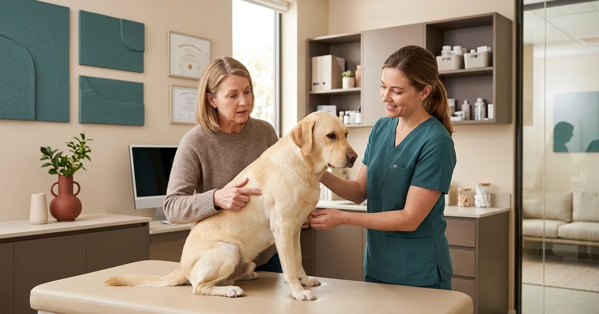
Nobody can tell whether your dog’s lump is cancerous by looking at it. Not you, not your regular vet, and not us. Benign and malignant masses can look and feel identical from the outside, which is why the reassurance you want has to come from a test rather than an opinion.
The good news is that the test is quick, cheap, and usually available the same day. The rest of this guide explains what we are looking for and what the common answers turn out to be.
Benign masses we see most often
Lipomas are benign fatty tumors, extremely common in middle-aged and older dogs, especially labradors and retrievers. They feel soft, they move freely under the skin, and they grow slowly. Most are harmless and need removal only when they get large enough to interfere with a leg or sit somewhere awkward.
Histiocytomas typically appear in young dogs as a small, round, raised, often hairless button. They can look alarming because they appear quickly and sometimes ulcerate. Many regress on their own within a couple of months.
Sebaceous adenomas are the small warty growths that accumulate on older dogs, particularly around the head and trunk. Benign, usually harmless, and worth removing when they bleed or catch on things.
The malignant one that matters most
Mast cell tumors are the most common malignant skin tumor in dogs, and they are the reason we take margins seriously. They are notorious for looking ordinary — a mast cell tumor can present as a soft, mobile lump that feels for all the world like a lipoma.
They also vary enormously in behaviour. Some are low-grade and cured by complete excision. Others are aggressive. A mast cell tumor removed with inadequate margins comes back, which is why identifying one before surgery changes the plan.
How we actually find out
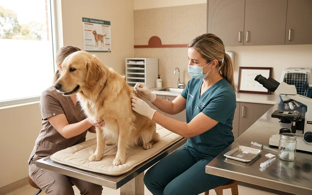
A fine-needle aspirate (FNA) is the first step. A thin needle draws a few cells from the mass, the sample goes onto a slide, and it can be examined quickly. It requires no sedation, it takes moments, and it is well tolerated by most dogs. An FNA will frequently identify a lipoma or a mast cell tumor with confidence, which is enough to plan the surgery properly.
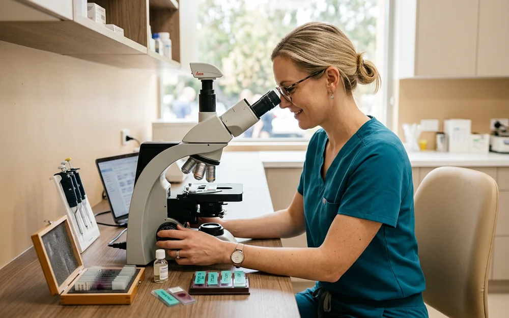
Histopathology is the definitive test and happens after removal. The whole mass goes to a laboratory, where a pathologist examines its architecture, grades it if malignant, and — because we ink the surgical margins before sending it — reports whether the tumor extended to the edge of the removed tissue. That last detail determines whether anything further is needed.
Warning signs that mean act now
Take a lump seriously if it is:
growing noticeably over weeks, firm rather than soft, fixed to the tissue underneath rather than moving freely, ulcerated or bleeding, changing colour, or accompanied by your dog being generally off.
Location matters too. A lump near a joint, an eye, or the mouth is harder to remove with adequate margins once it grows, so the surgical window is narrower.
What to do next
If you have found a lump on your dog, the practical move is an evaluation with an aspirate rather than weeks of anxious watching. It gives you an answer quickly, and if the answer is benign you can stop worrying with something more solid than hope.
If it is not benign, early removal with proper margins is the single biggest factor in the outcome. Book a mass evaluation and we will tell you plainly what we find, what we recommend, and what it costs — including when the recommendation is simply to keep an eye on it.