The four reasons a spleen comes out
Spleen removal is performed for a small number of clear indications, and understanding which one applies to your dog shapes both the urgency and the conversation about outcome. Our splenectomy service handles all four.
A splenic mass. A growth on or within the spleen, found either on an ultrasound performed for another reason or after the dog becomes unwell.
A splenic hematoma. A blood-filled swelling within the spleen, often the benign explanation for what looked like a mass on imaging.
Acute rupture. Usually after trauma such as a road accident or a heavy fall, but sometimes a mass that has bled spontaneously.
Hemoperitoneum. Free blood in the abdominal cavity from a bleeding spleen. This is the emergency presentation.
Emergency or planned?
The emergency presentation is dramatic and unmistakable in hindsight. A dog — often an older large-breed dog — collapses suddenly. The gums are pale. They are weak, and the abdomen may look distended. This is a bleeding spleen filling the abdomen. It is time-critical and needs stabilisation and surgery without delay.
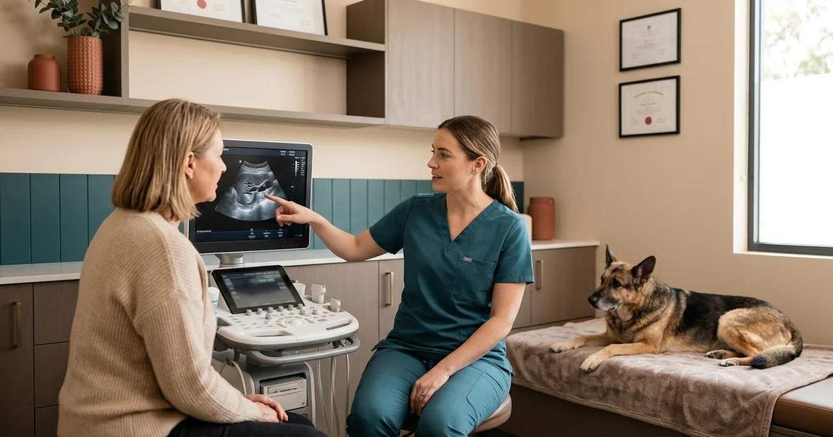
The incidental finding is the better version. A splenic mass is picked up on an ultrasound performed for something entirely unrelated, in a dog who seems completely well. This allows for staging, planning, and a proper conversation before anyone books a theatre.
The fifty-fifty conversation
About half of canine splenic masses are benign. No scan can tell you which half your dog is in.
That statistic does more work than any other in this conversation. Owners facing a splenic mass diagnosis frequently assume the worst and ask whether surgery is worth putting their dog through.
The honest answer is that surgery is simultaneously the treatment and the diagnosis. Only histopathology of the removed spleen tells you what the mass actually was. Declining surgery does not avoid a cancer diagnosis; it avoids ever having one, while leaving in place a mass that may bleed catastrophically.
What we do before operating
Abdominal ultrasound characterises the splenic lesion and examines the liver, which is the most common site of spread if the mass is a hemangiosarcoma.
Thoracic radiographs look for pulmonary metastasis.
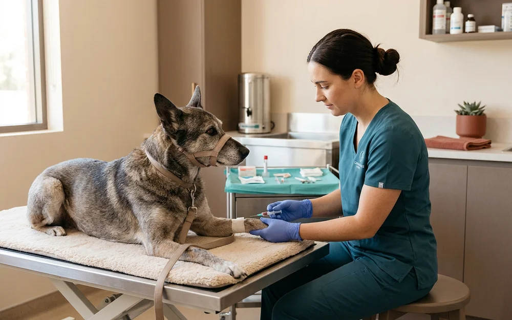
Bloodwork establishes a baseline packed cell volume so ongoing blood loss can be measured against it, and checks clotting where there has been bleeding.
Where a dog is actively bleeding, stabilisation with intravenous fluids — and a transfusion where the packed cell volume has fallen far enough — comes before anesthesia.
Age is not the deciding factor
Splenic disease is largely a disease of older dogs, so age alone cannot rule surgery out. What matters is organ function and cardiac status, both assessed before we proceed.
A bleeding spleen left in place is not the conservative option. It is a mass that will bleed again, and the next bleed may not be survivable.
What recovery looks like
Most dogs go home within a day or two, with two weeks of restricted activity while the abdominal incision heals. We monitor packed cell volume and lactate through the early recovery because those numbers reveal ongoing bleeding before it becomes visible.
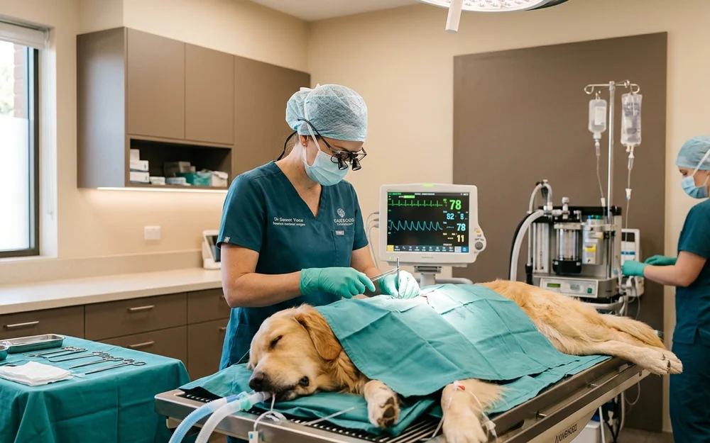
Continuous ECG monitoring runs during and after surgery, because splenic cases are associated with ventricular premature contractions. Most are self-limiting; a proportion need treatment.
Histopathology typically returns within a week. Whatever it says, we will explain it plainly — including, if the news is not good, what the realistic picture is rather than a version designed to make the conversation easier.