What holds a broken bone still
A fracture heals when the bone ends are held in the right position, without movement, for long enough. Everything in fracture surgery serves that one requirement.
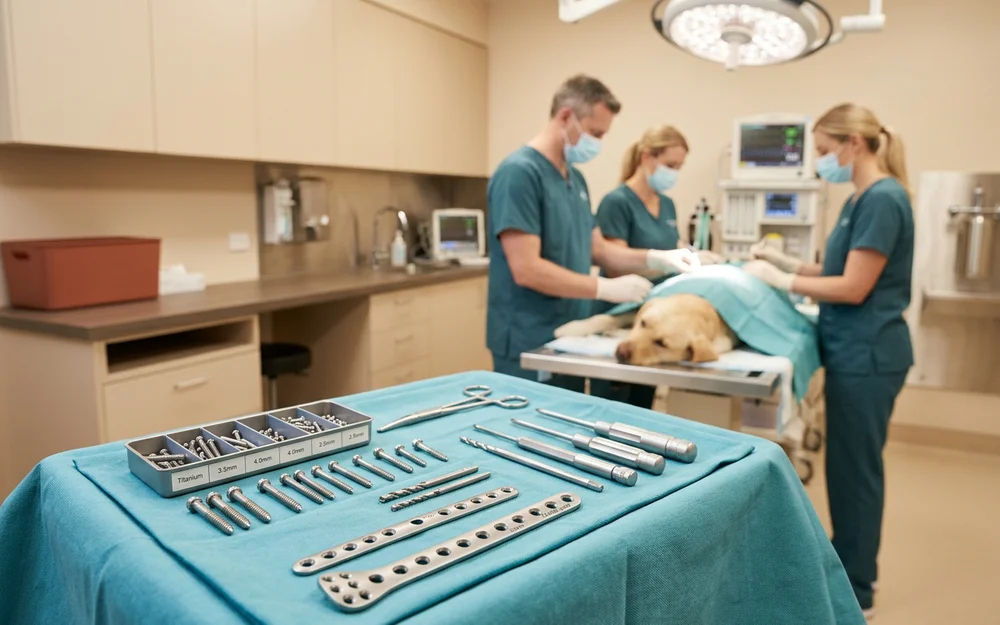
Our fracture repair service uses three main approaches, chosen by the fracture pattern rather than by preference.
Dynamic compression plating (DCP and LC-DCP). A metal plate is contoured to the bone and secured with stainless steel cortical screws. The plate design allows the screws to compress the fracture fragments together as they are tightened, which is exactly the environment bone prefers for healing. Plates give rigid stability and suit long-bone fractures under load.
Intramedullary (IM) pinning. A pin is driven down the marrow cavity, along the axis of the bone. It resists bending very effectively and rotation hardly at all, which is why an IM pin is usually combined with K-wires or a cerclage wire to control rotational forces.
Kirschner wires (K-wires). Fine wires used in smaller bones, in young dogs, or alongside a pin to hold individual fragments in position.
External fixation. Pins pass through the skin into the bone and connect to an external frame. Because no hardware is implanted at the fracture site itself, this is the approach of choice for open or heavily contaminated fractures where implant infection is a genuine risk.
How bone actually heals
Bone does not heal like a cut. It goes through defined stages, and each one takes time that cannot be shortened.
Inflammation (days 1 to 7). A blood clot forms at the fracture site and inflammatory cells clear debris. The limb is at its most painful in this period.
Soft callus (weeks 1 to 3). A bridge of cartilage and fibrous tissue forms across the fracture. It is holding the bone ends together but it is not yet strong.
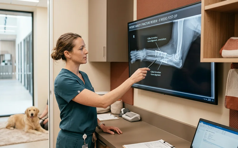
Hard callus (weeks 3 to 8). The soft callus mineralises into woven bone. This is where callus formation becomes visible on radiographs, and it is the stage we are looking for before lifting restrictions.
Remodelling (months). The woven bone is gradually reorganised into normal bone architecture along the lines of stress. This continues long after your dog is walking normally.
Why recheck radiographs decide everything
How well your dog is walking tells you nothing useful about how well the bone is healing.
Dogs are enthusiastic weight-bearers. A dog three weeks into a twelve-week healing timeline will happily trot on a leg held together entirely by hardware and soft callus, and will interpret any relaxation of the rules as permission.
Orthogonal recheck radiographs — two views at right angles — show the actual state of the callus. Restrictions come off when the images say so.
The recovery nobody warns you about
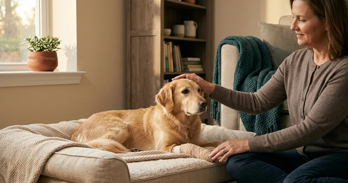
Strict confinement is genuinely difficult, and it is the part of fracture repair that owners underestimate every single time.
- Crate or single-room rest
- Lead-only toilet breaks, short
- No stairs, no furniture, no car jumps
- No off-lead running until cleared on radiographs
- No play with other dogs
Non-union prevention is almost entirely about this. A fracture that fails to knit is usually a fracture that was not held still enough, and the movement almost always came from a dog who felt fine.
Six to twelve weeks of enforced boredom is a long time. It is also the difference between a leg that works normally for the next decade and one that does not.
Before any of it
If your dog has been hit by a car, go to your nearest emergency hospital first. Chest injury, abdominal bleeding, and shock kill in the first hours; the leg does not. Once your dog is stable, we can plan the repair.